Instrument Reservation - Stratocore
Check out our new Hitachi FlexSEM 1000-II scanning electron microscope at the end of the page under "Electron Microscopes".
Advanced Fluorescence Microscopes
Confocal Microscopes
Nikon A1R-Ti2 confocal system
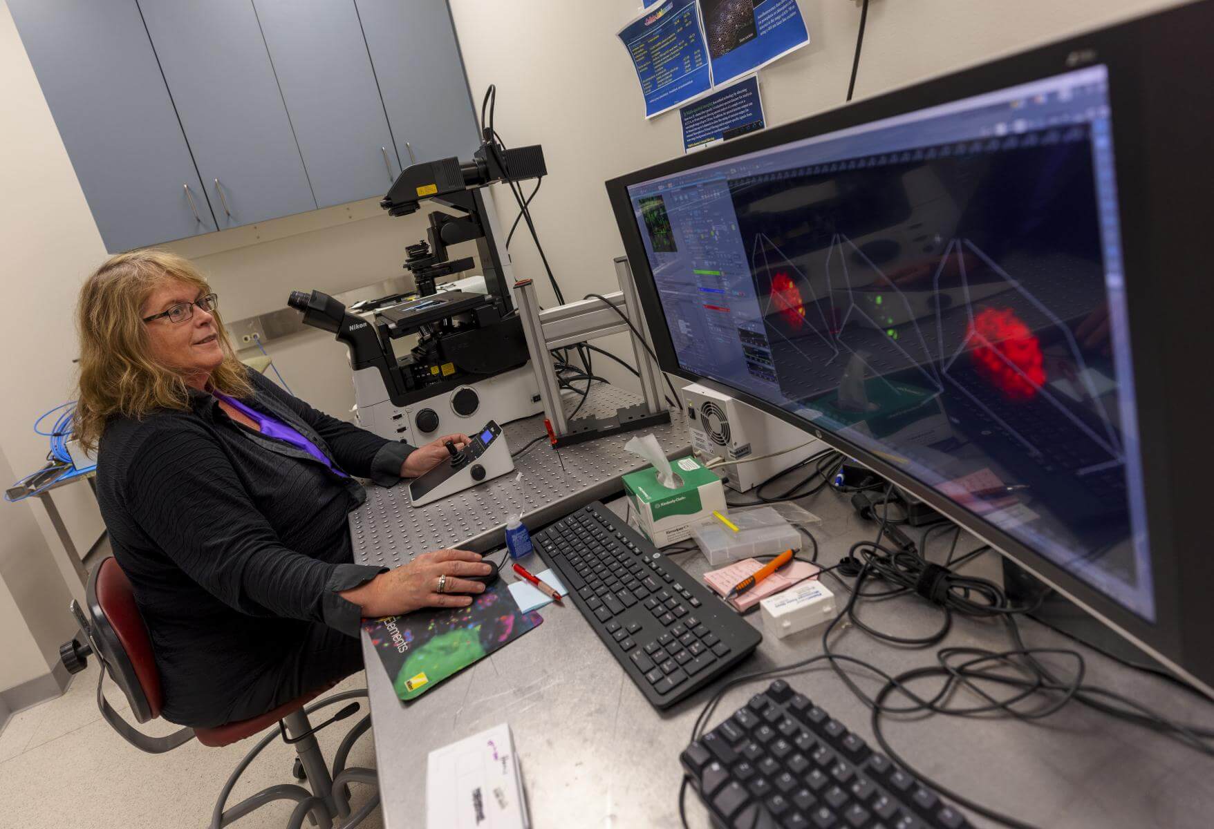
| This confocal is best for live cell imaging analysis using cultured cells (in culture dishes or well plates). The cutting-edge imaging system includes a hybrid resonant dual scanner unit for ultrafast photo-activation imaging and integrated six-solid-state laser package (with 405nm, 440nm, 488nm, 514nm, 561nm, and 640nm laser lines). It also has an ultra-sensitive GaAsp hybrid 4+1 channel detector system and an advanced inverted microscope with a motorized stage and a stage-top incubator unit, which can be preprogramed to image samples using glass slides, culture dishes, or multiwall (6-96) plates. |
Nikon A1-NiE confocal system
This confocal is integrated with an upright fluorescence microscope (Ni-E) with a motorized stage, which allows the user to carry out image analysis of large samples. This confocal also has an integrated six-solid-state laser package (with 405nm, 440nm, 488nm, 514nm, 561nm, and 640nm laser lines), and the NIS-Elements Confocal image acquisition and analysis program. Note: We also have a computer workstation for analysis of image data obtained using Nikon NIS-Element | 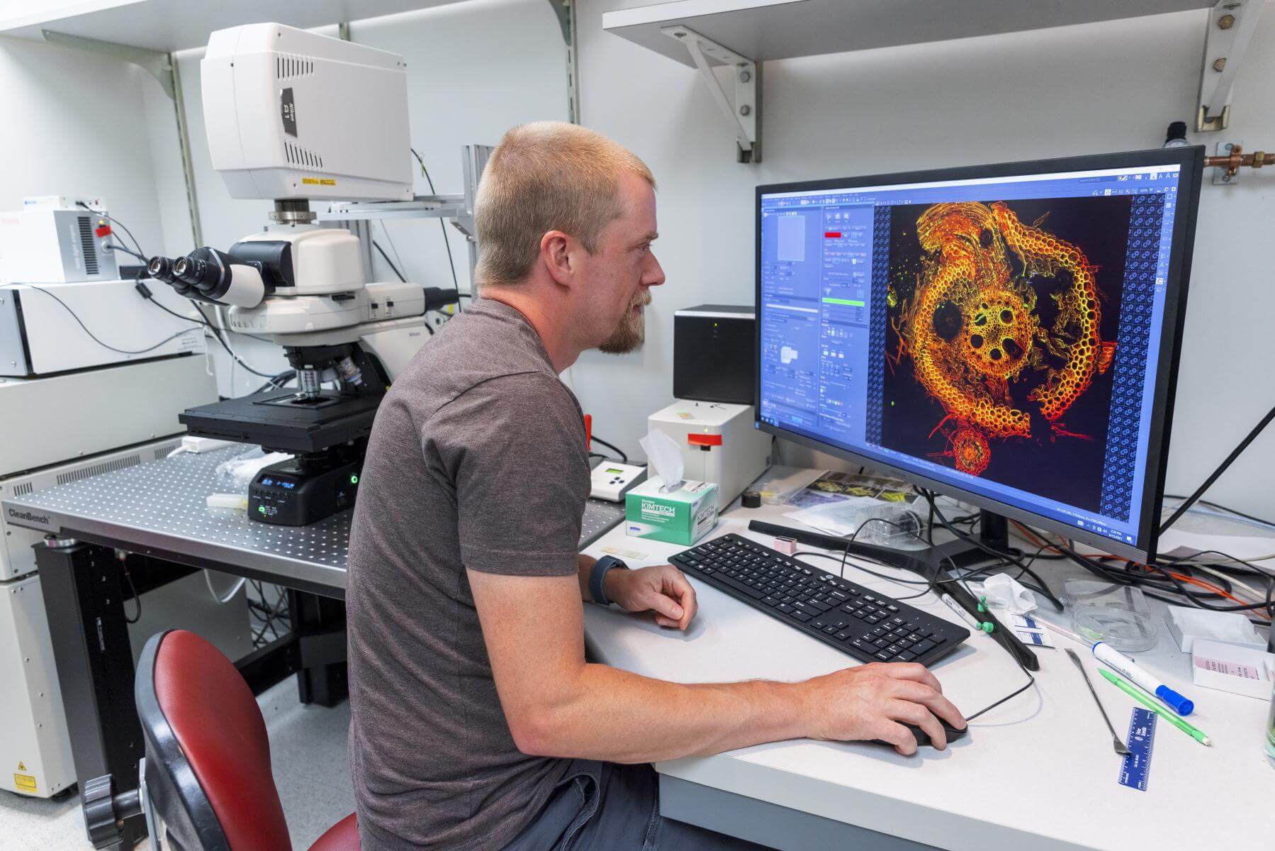
|
Nikon Ti-2 Inverted Fluorescence Microscope
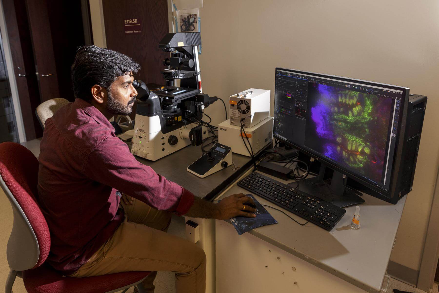
| It is equipped with automated stage and due digital cameras (Qi-@ mono with high sensitivity for low light/fluorescence, and F1-2 for colorimetric image). This system allows researchers to collect images using regular slides and dishes of cultured cells (or well plates) under sterile conditions for live cells imaging. The Ti-2 system has specific set of filter cubes (6 cube positions), including, DAPI, CFP, GFP, YFP, CY3/DsRed/RFP, mCherry/Texas Red, CY5, and CY7, allowing sequential image collections of samples with up to five fluorescent tags (including Cy7) with proper spectrum separation. |
Upright Fluorescence Microscope
| (Olympus AX70) with digital camera. | 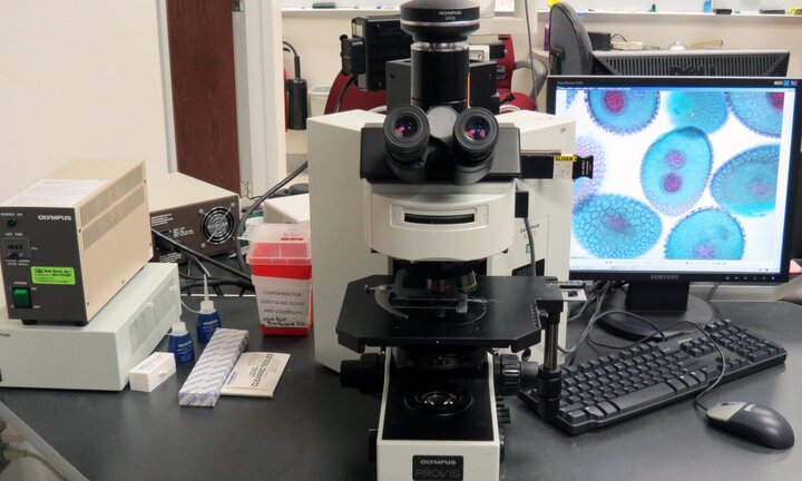
|
Stereo Fluorescence Microscope Nikon SMZ25 with DS-Ri2 Camera
| Stereo fluorescent microscope with dissecting illumination and epi-fluorescence illumination, 3 LED Light cubes (GFP (488), DS Red (560) and Long Pass (GFP and DS Red), 2 Lenses (.5X with Optical Zoom from .32 to 7.88) and (1X with Optical Zoom from .63 to 15.75) which allows users to screen fluorescent samples or capture images for phenotypic selections. |
EVOS® FL Auto Cell Imaging System
| EVOS® FL Auto Cell Imaging System with an automated microscopic stage (computer controlled X-Y-Z movement), dual cameras, selectable excitation/emission filters from 5 LED Light Cubes (DAPI, GFP, RFP, Texas Red, and Cy5), and 3x optical zoom over different lenses (4x, 10x, 20x, 40x, 60x, and 100x). This system is user friendly and suitable for single or multi-labeling fluorescence or colorimetric image acquisitions from sections or live cells using single culture dish, 6, 12, or 24-well culture plates, or 96 well ELISA plate. | 
|
Electron Microscopes
Transmission Electron Microscope
| Hitachi HT-7800 TEM is an advanced electron microscope for a) ultrastructural analysis on ultrathin sections of samples, b) assay of conductive particles, and c) examination of negative stained microbial particles (such as virus) and nanoparticles. A side-mount high-resolution CCD camera is installed with this system. Magnification range: 50x to 200,000x using high contrast mode at 80 KV; Up to 600,000x under the high-resolution mode. |
Hitachi FlexSEM 1000-II scanning electron microscope
The SEM (installed on Feb 12 2025) combines innovative technological features with an intuitive interface, to deliver adaptability and flexibility in a powerful, automated, lab-friendly package with the following newer capabilities and specs: a) Allow users to use uncoated biological samples; b) Ultra-Variable Pressure (UVD) Detector; c) Uni-Bias (Even emission across entire voltage range); d)Integrated Oxford EDS (The X-ray system-EDS can operate on the same PC; e)Variable pressure operation (6 to 100Pa range); f) Dual=operation mode (user-friendly, Easy Mode beginning users or student to get great images. Advanced Mode for experts); g) large sample size allowed: up to 80 mm diameter of surface area and up to 40 mm of sample thickness; h) 3-axis (X/Y/R) Motor Driven Stage: X:50mm, Y:40mm, Z:5-33mm, Rotation: 360 degrees, Tilt: -15 to +90 degrees; Magnification Range: 16x to 800,000x (display); Accelerating Voltage Range: 0.3kV to 20kV | 
|
Other equipment and services
Cell culture unit with a tissue culture hood (Biosafety Cabinet)
This cell culture unit was designed for live cell image analysis so that users do not have to transport live cells to the core and can do different cell treatments within the imaging core.
Sputter coating systems
- Techniques Sputter is used for larger sputtering particles and thicker coating for large samples such as rocks, leaves, and food products.
- Denton Desk V Sputter is used for small sputtering particles when using high-resolution topographic analyses of cells and biological tissues at high magnification.
Microtomes and sectioning services
- Leica Cryostat is used for the preparation of frozen sections for animal or plant tissues for immunofluorescence or histochemical localization analyses of specific molecules or proteins of interest.
- Leica Rotary Microtome is used for paraffin sectioning for thin sections for histological and immunochemical image analyses.
- Leica EM UC7 Ultramicrotome is used for the preparation of semi- and ultra-thin sections of biological samples, including animal/plant cells or tissues and microorganisms.
Other Services
The core has staining tools for immunofluorescence labeling for confocal Microscopy and immunogold labeling for electron microscopy. We can also provide training for users to carry out such experiments in their own labs.