Content
OUR CORE FACILITIES
When the Center for Biotechnology was formed in 1988, one of the priorities was to develop state-of-the-art core research facilities that would provide the technical expertise and equipment needs of faculty, staff, students and Nebraska-based businesses on a service basis to conduct research in the field of biotechnology. These facilities continue to function to provide access on the University campus to state-of-the-art scientific expertise and equipment that are too costly for individual scientists or businesses to have in the laboratories they direct. Another primary function of the facilities is to provide opportunities for educating students about the modern techniques used in the field of biotechnology.
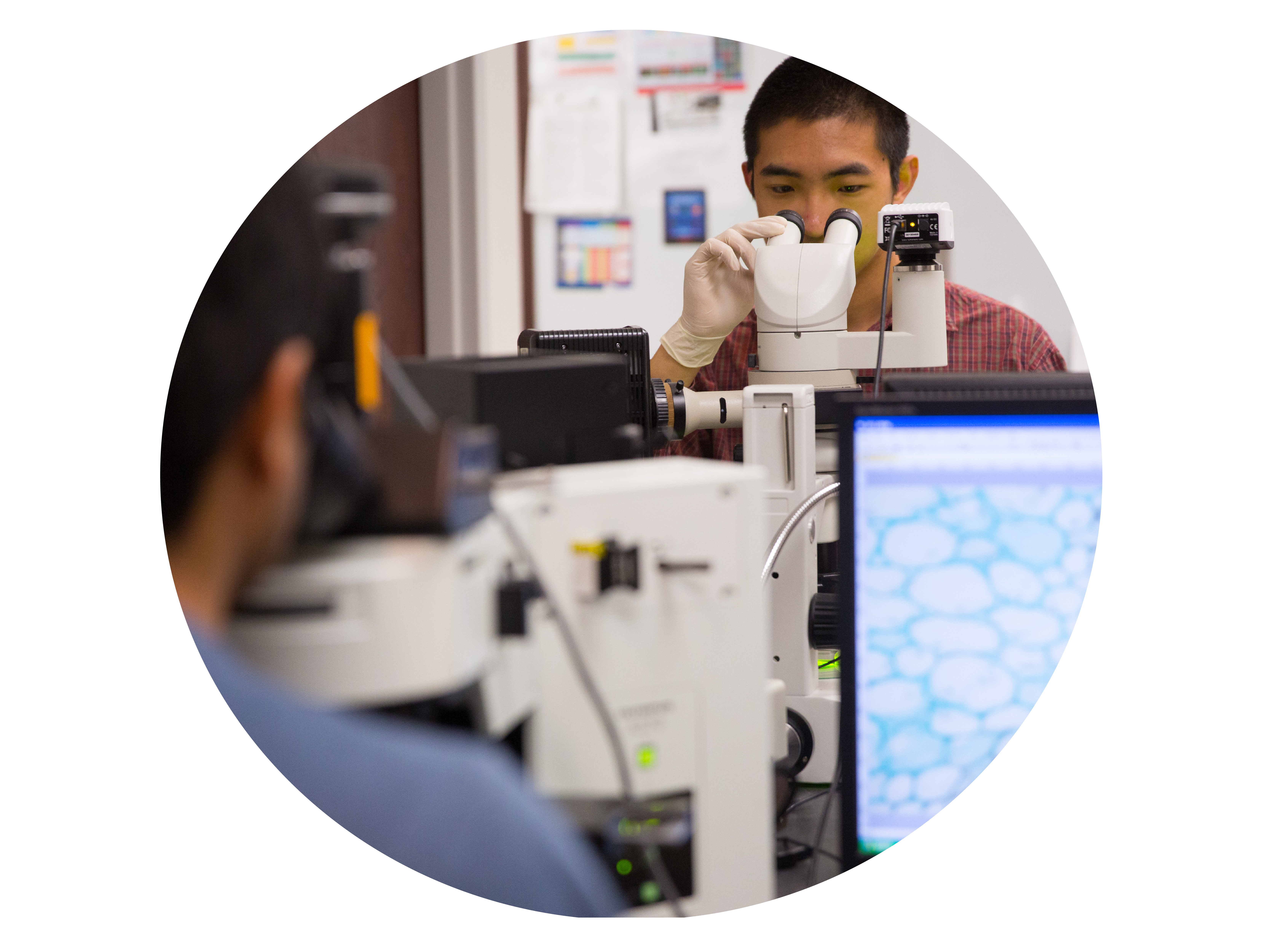
MICROSCOPY
Located on the first floor of the George W. Beadle building, the Microscopy Core Research Facility has state of the art imaging systems including light/fluorescence microscopes, confocal laser scanning microscopes and electron microscopes.
Learn more

FLOW CYTOMETRY
The Flow Cytometry Core Facility provides flow cytometry services to investigators on and off the University of Nebraska-Lincoln campus on a fee for service basis. The core provides comprehensive data collection, data interpretation and education in flow cytometry.
Learn more
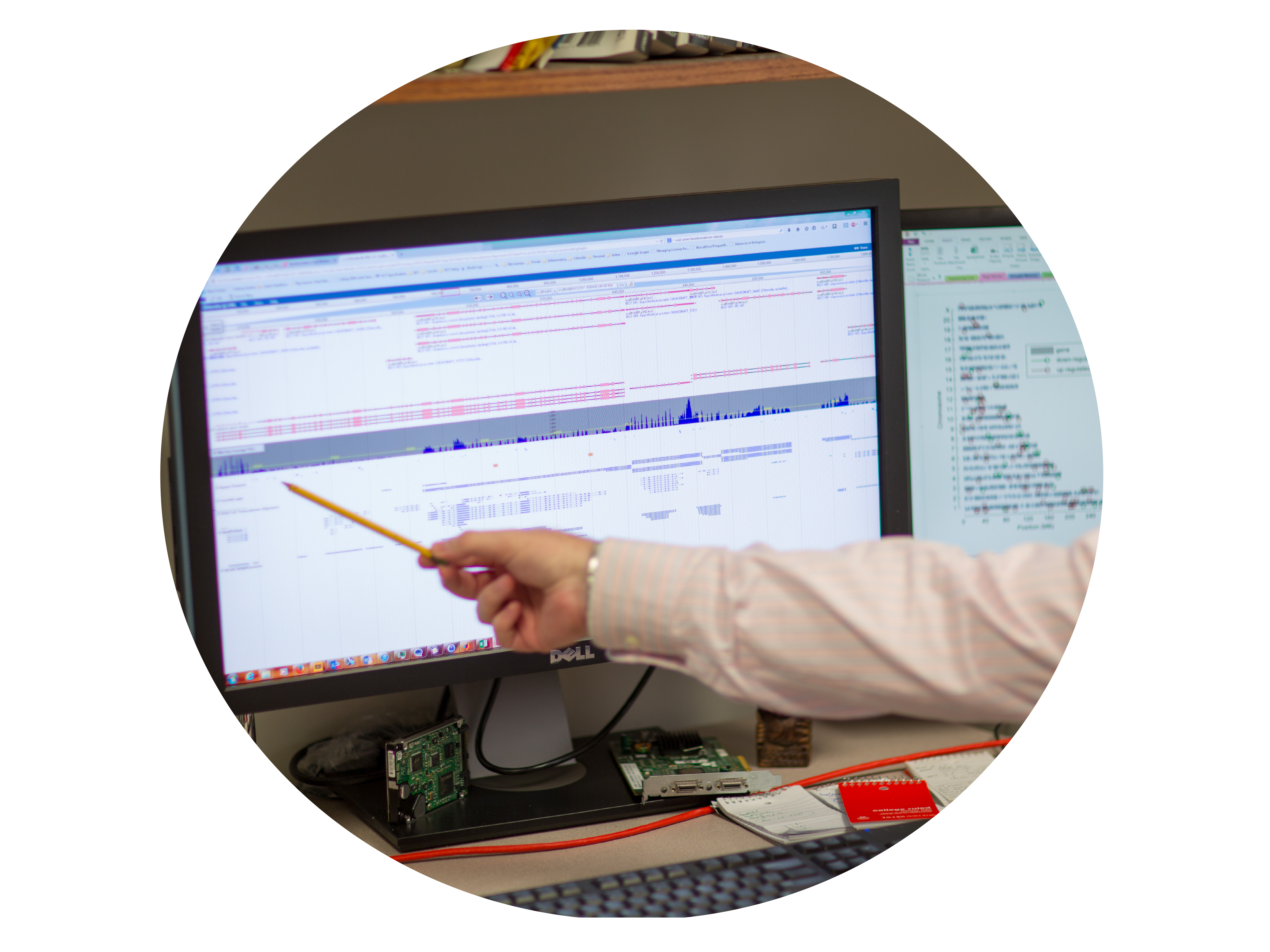
BIOINFORMATICS
The Bioinformatics Core Research Facility offers education, analysis, and computational services in the area of bioinformatics and computational biology.
Learn more
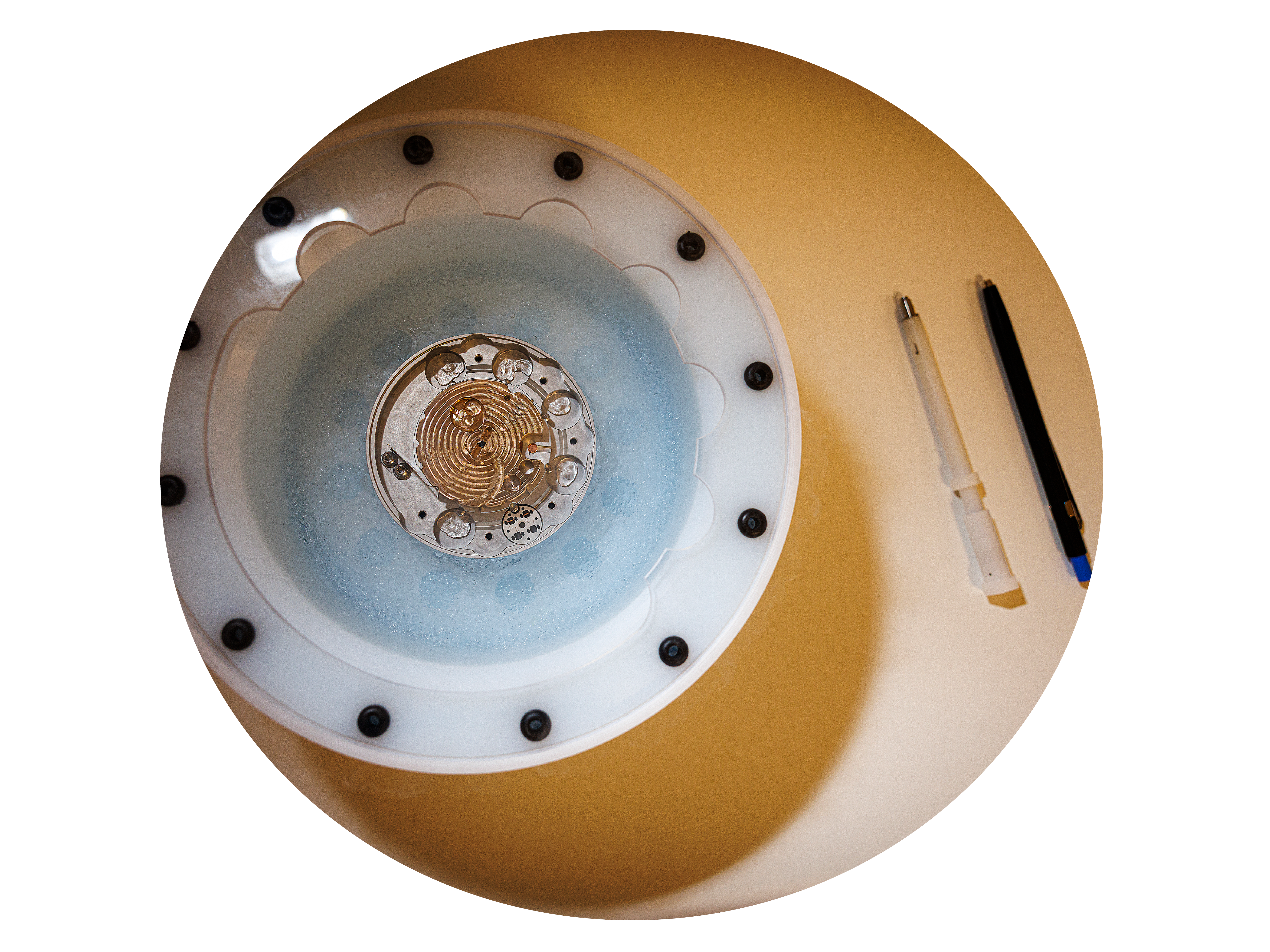
CRYOEM
Cryo-EM is a structural biology technique that has progressed exponentially in the last few years. It has an unparalleled capacity to provide near atomic-resolution structures of biological macromolecules. It is also uniquely suited to provide information about molecular rearrangements and interactions that are often essential to the function and mechanism of macromolecular complexes involved in a variety of cellular processes.
Learn more
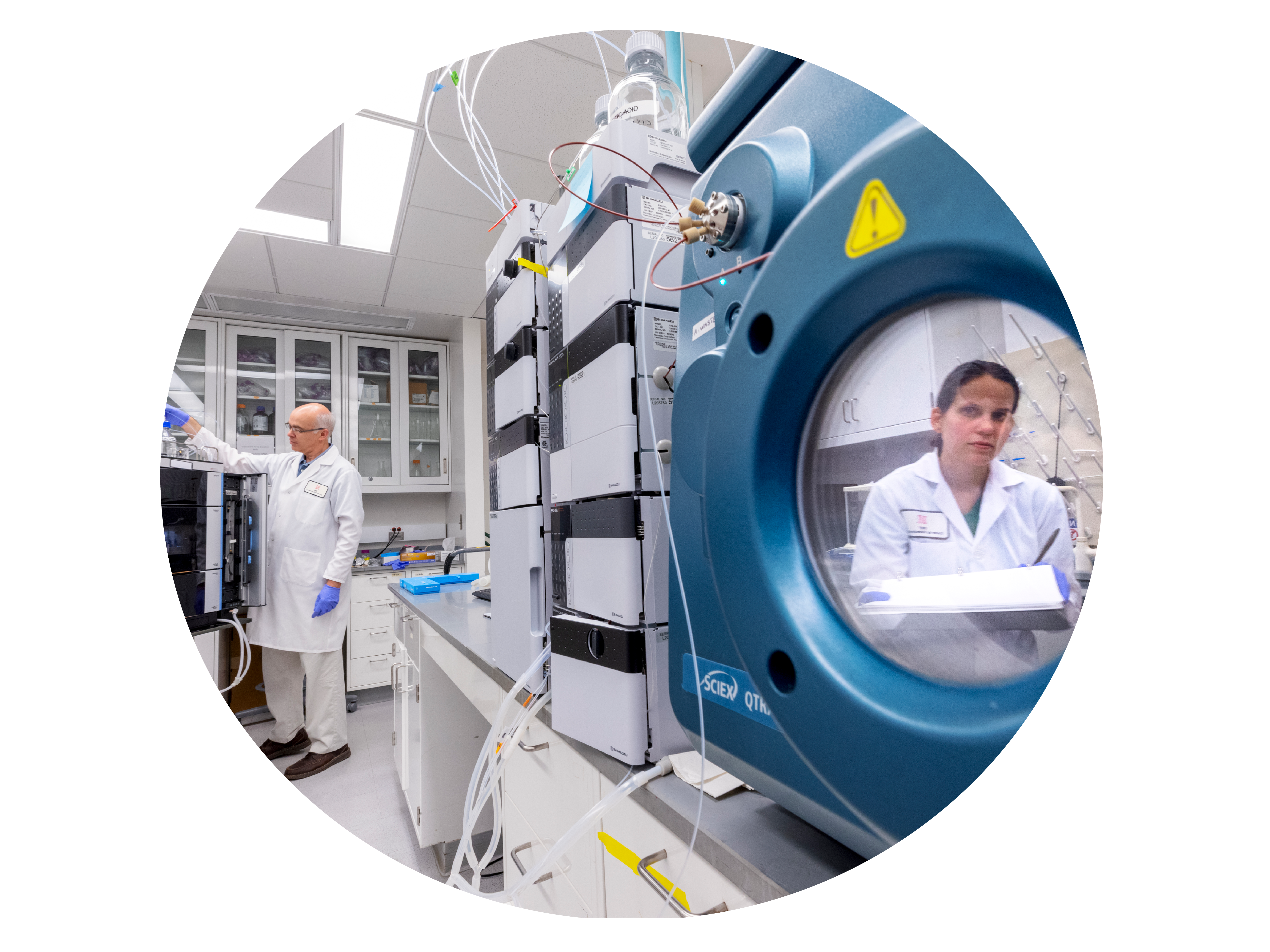
PROTEOMICS AND METABOLOMICS
Newly equipped facility with the latest state-of-the-art mass spectrometers and bioinformatics tools. Suitable for addressing complex proteomics and metabolomics questions at multiple levels.
Learn more
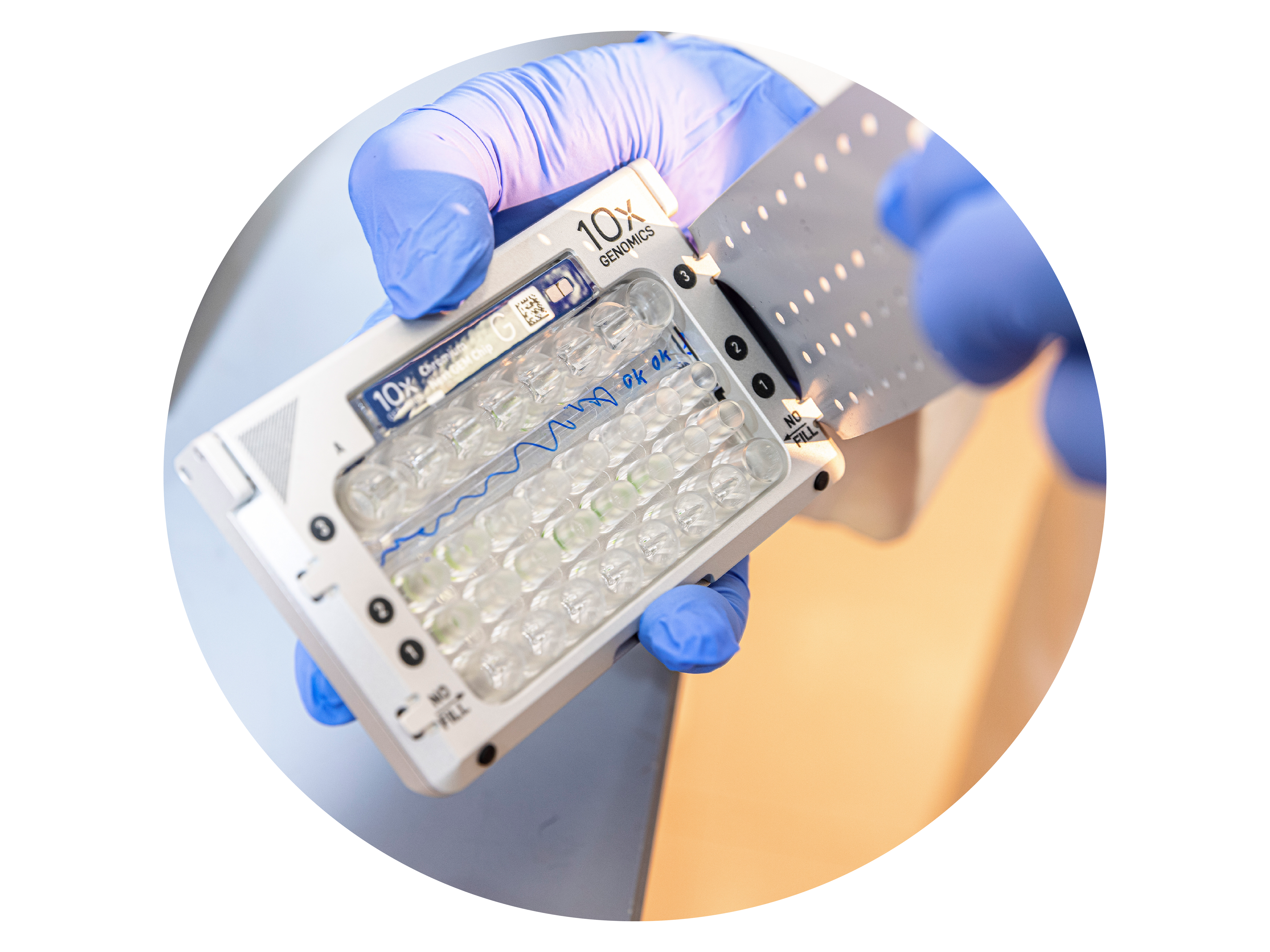
SINGLE CELL GENOMICS
The Single Cell Genomics Research Facility
Learn more